Radiology & Imaging37 Videos


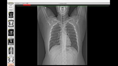
How I Read a Chest CT
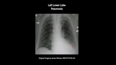
What are some common uses of the procedure? Doctors use chest CT to: examine abnormalities found on chest x-rays. help diagnose the causes of signs or symptoms of chest disease, such as cough, shortness of breath, chest pain, or fever. detect and evaluate the extent of tumors that arise in the chest, or tumors that […]
How to Read a CT Scan of the Head
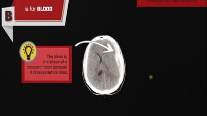
What is a head CT? Computed tomography, more commonly known as a CT or CAT scan, is a diagnostic medical imaging test. Like traditional x-rays, it produces multiple images or pictures of the inside of the body. A CT scan generates images that can be reformatted in multiple planes. It can even generate three-dimensional images. […]
What Happens During a CT Lung Scan
computed tomography of the chest Computed tomography (CT) is an imaging technique that has revolutionized medical imaging. It is widely available, fast, and provides a detailed view of the internal organs and structures. Helical CT is most common, but conventional, axial, step-and-shoot CT is used for thin section high-resolution CT scanning of the lungs, coronary […]

