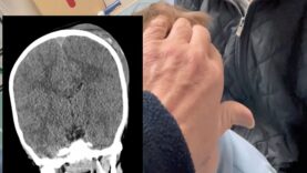
Describing the typical appearance of skull fractures as seen on computed tomography (CT) imaging Mediube Subscribe Subscribed 3 1173 videos 0% 7 Views 0 Likes 2024-06-30 Head & Neck By Mediube 0 Comments (بازدید 7 بار, بازدیدهای امروز 1 ) Show more PREV Understanding Subdural Hematoma Mediube 2024-06-29 NEXT Tripod Other Cheekbone Facial Fractures Mediube 2024-07-14 You might be interested in 0 BONES OF THE SKULL LEARN IN 4 MINUTES Mediube 2024-08-05 0 Mandible structure and bony landmarks Mediube 2025-02-08 0 Skull Anatomy Mediube 2024-08-08 0 Head and Neck Assessment Nursing Mediube 2024-03-27 0 Neck Anatomy – Organisation of the Neck Mediube 2024-07-31 0 Intracranial Hemorrhage- Epidural-Subdural-Subarachnoid overview Mediube 2024-06-24 0 Brain anatomy function Mediube 2024-08-05 0 Tripod Other Cheekbone Facial Fractures Mediube 2024-07-14 1 Subdural Hematoma VS Epidural Hematoma – Intracranial Hemorrhages Mediube 2024-06-29 0 Triangles Subdivisions of the Neck – Head and Neck Anatomy Mediube 2024-08-03 0 Skull Fracture Emergency in a 4-Year-Old Boy Mediube 2024-06-29 1 CT Head Interpretation for Beginners – OSCE Guide – UKMLA – CPSA Mediube 2024-05-27 LEAVE YOUR COMMENT Cancel replyYour email address will not be published. Required fields are marked * Save my name, email, and website in this browser for the next time I comment. Δ


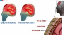

![Subdural vs Epidural Hematoma-Hemorrhage [CT Scan Findings]](https://mediube.com/wp-content/uploads/2024/06/Subdural-vs-Epidural-Hematoma-Hemorrhage-CT-Scan-Findings-205x115.jpg)