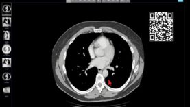
Computed tomography, more commonly known as a CT or CAT scan, is a diagnostic medical imaging test. Like traditional x-rays, it produces multiple images or pictures of the inside of the body.
A CT scan generates images that can be reformatted in multiple planes. It can even generate three-dimensional images. Your doctor can review these images on a computer monitor, print them on film or via a 3D printer, or transfer them to a CD or DVD.
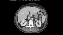
CT images of internal organs, bones, soft tissue, and blood vessels provide greater detail than traditional x-rays. This is especially true for soft tissues and blood vessels.
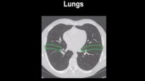
Doctors can use a variety of techniques to lower the amount of radiation needed to perform a chest CT scan. These techniques include adjusting the radiation dose based on patient size and new software technology. A low-dose chest CT produces images of enough quality to detect many lung diseases and abnormalities using much less radiation than a conventional chest CT scan. In some cases, doctors may lower the dose by 65 percent or more. Doctors routinely use low-dose chest CT to evaluate acquired and congenital lung abnormalities. These include pneumonia, interstitial lung disease, and tumors. There is ongoing research to lower radiation doses even further. Your radiologist will decide the proper settings for your scan depending on your medical issue and what information the CT scan needs to obtain. If your child is to have a CT scan, the doctor will use low-dose pediatric settings.